Canine Hip Dysplasia (CHD), by definition, means that the hip is anatomically abnormal. The hip is a ball and socket joint, and hip dysplasia occurs when the ball does not fit tightly into the socket. A loose fit is known as joint laxity. This joint laxity is what is responsible for the development of clinical signs in the dog, as the ball of the femur (the femoral head) moves around too much inside the socket (the acetabulum). This movement results in the wearing down of the femoral head, damage to the acetabulum, and bony changes as the joint tries to stabilise itself.
This condition is present when the dog is a puppy. However, the dog usually won’t show symptoms until around 6-9 months as the ligaments and connective tissue around the joint start to become painful due to stretching. This is known as Juvenile Onset CHD. Some dogs won’t show signs until even later, after 2 years of age, when osteoarthritis is starting to develop and resulting in pain. This is known as Adult Onset CHD.
CHD occurs most commonly in large breed dogs. However, medium and small breed dogs can be affected as well. Some common breeds affected include Labrador Retrievers, German Shepherds, French Bulldogs, English Bulldogs, Great Danes, Rottweilers and Golden Retrievers, to name a few.
There are multiple factors that lead to the development of CHD. However, genetics are the most significant risk factor. Studies have also shown that rapid weight gain/growth, especially during puppyhood, also contributes to the development of CHD.
How is it Diagnosed?
Diagnosis is based on several components:
- Thorough physical examination, assessing the dog’s range of motion in each leg, and checking for a pain response. Sometimes swelling may be felt at the affected joint. A common finding in dogs with hip dysplasia is a reduction in muscle on the affected legs.
- The Ortolani test: This is a test performed under sedation or anaesthesia to determine the laxity of the joint. The hip is moved in such a way that if the joint is loose, the femoral head will be heard and felt to ‘clunk’ in and out of the socket. In puppies or non-painful dogs, this may be checked without sedation.
- Radiographs: Radiographs can give an indication of how loose the joint is, how much of the ball sits inside the socket, and show us how severe the development of osteoarthritis is. This will definitively diagnose hip dysplasia. These can also be used to determine what surgeries may be an option for the dog.
What can be done?
Non-surgical management: Most dogs who are diagnosed with hip dysplasia as adults are managed medically. The goal is to make the dog feel more comfortable and delay the progression of further osteoarthritis. This includes weight management, pain relief, laser therapy, acupuncture, and supplements/injections to improve the functionality of the joint. More information can be found in the attached ‘Joint Health – Managing Osteoarthritis’ sheet.
Surgical management: There are two main surgical options for adult dogs with hip dysplasia and osteoarthritis:
- Femoral Head and Neck Ostectomy (FHNO): This procedure removes the head of the femur (the ball of the joint), which reduces the pain associated with stretching of the ligaments and osteoarthritis. The limb then slowly forms a ‘false joint’ of fibrotic tissue around the area to allow use of the limb. Dogs will not regain complete normal function, however it will help greatly to alleviate the pain. This procedure is best for dogs under 30kg, but may be considered in larger dogs depending on the severity of the hip dysplasia in both limbs.
- Total Hip Replacement (THR): This is a specialist procedure where the hip joint is replaced with an artificial joint to allow a more natural range of motion and limb function. This produces are more normal functioning limb, so is more suited to dogs who work, perform in agility, or other strenuous activities. As the hip joint has been removed, the pain associated with the laxity and osteoarthritis is removed.
Hip Dysplasia + Osteoarthritis: The following images are a comparison of a dog with hip dysplasia to one with normal hips.
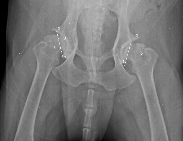
The first image shows:
- Bone spur formation (osteophytes)
- Thickening of the femoral neck
- Thickening of the socket bone due to increased wear.
The white line highlights how much of the femoral head is sitting inside the socket. It is far less than 50% of the femoral head, demonstrating hip dysplasia.
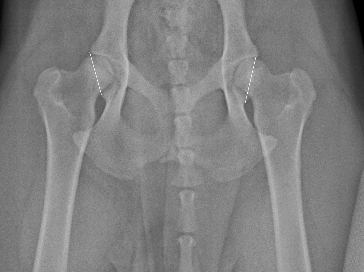
The second image shows:
- No sharp bony spurs
- Normal femoral necks with a ‘waist’
- No thickened bone in the socket
The white line highlights how much femoral head is sitting inside the socket. It is greater than 50% of the femoral head, which is ideal.
For more information on how to manage osteoarthritis in dogs, please see our article Joint Health In Dogs.